
 TH
TH
A CASE REPORT:CLINICAL APPROACH OF BLINDNESS FOLLOWING HYDROCEPHALUS IN DOG
Kriengkrai Chatthakulchai 1, Onpraween Thangthanasirikul 2
1, 2 Eye clinic, Thonglor Pet Hospital Bangkok, Thailand
Keyword: Hydrocephalus, Transcranial ultrasound, Height of lateral ventricle, Ventricle-mantle ratio
Introduction
Acute blindness in dogs may occur with many ophthalmic diseases (retinal disease, optic nerve disease) and central nervous system disease (intracranial disease). Blind dogs are divided into two groups; Central blindness and peripheral blindness, by PLR. Central blindness could be suspected if PLR responds normally, while peripheral blindness is described by the abnormal PLR in the multifocal lesion of ocular media, retina, optic disc or optic chiasm. Lesion influencing central visual pathway e.g. hydrocephalus, GME causes vision loss with normal PLR and result in other clinical signs of forebrain disease such as decreased consciousness, seizure or circling. Hydrocephalus is described as; the abnormal accumulation of cerebrospinal fluid in the brain; is a common cause of central blindness in dog. Toy breeds and dogs with dome-shaped foreheads are predisposing breeds of hydrocephalus especially Chihuahua, Pomeranian and Maltese. The congenital hydrocephalus is characterized by appearance of bilateral ventrolateral strabismus.
Material and method
A- eight months old, female, Chihuahua dog developed blindness within the week. She would walk slowly and bumps into things around the house. Clinical approach started from a complete eye examination.
Table1: ophthalmic examination
| Parameter | Left eye | Right eye |
| IOP (mmHg) | 12 | 14 |
| Menace response | negative | negative |
| Maze test | negative | negative |
| Dazzle reflex | positive | positive |
| Pupil size | dilate | dilate |
| Direct PLR | positive | positive |
| Indirect PLR | positive | positive |
IOP: intraocular pressure, PLR pupillary light reflex
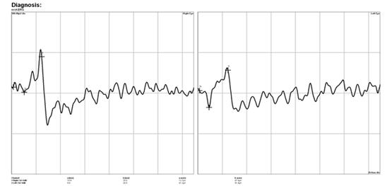
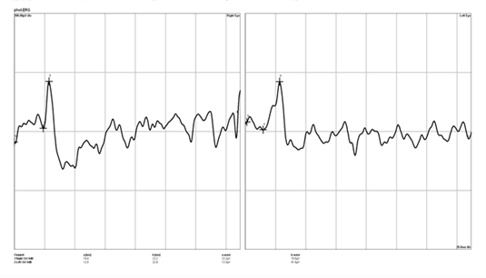
This dog developed bilateral blindness with normal pupillary light reflex (both direct PLR and indirect PLR) thus, central blindness was suspected. Next step, ERG (Electroretinography) was used to evaluate retinal function and rule out peripheral blindness.
Electroretinography (ERG)
ERG (RETIport ERG-An-vision™) revealed that the amplitude of A-wave and B-wave was in normal range so intracranial disease was a likely cause of central blindness.
Next step, an ultrasound (Hitachi Healthcare’s Prosound Alpha 7™) was performed to measure the height of lateral ventricle and ventricle-mantle ratio to check the ventriculomegaly condition.
Ultrasonography
Transcranial ultrasound is an invasive, without anesthesia, diagnostic technique of canine hydrocephalus. Hitachi Healthcare’s Prosound Alpha 7™ measures the height of lateral ventricle (dorsoventral dimension) and ventricle-mantle ratio (ratio of lateral ventricle height to the thickness of cerebral mantle).
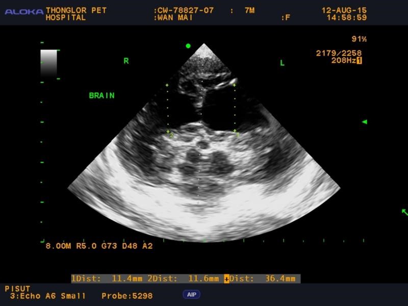
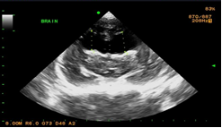
Ultrasound result: The ventricle-mantle ratio is abnormal so ventriculomegaly is indicated
| Parameter | Result | Normal range |
| The height of lateral ventricle | 0.11 cm | 0.04-0.35cm |
| Ventricle and mantle ratio | 0.11 cm / 0.36 cm = 0.31 | 0.25 |
Diagnosis: Hydrocephalus was diagnosed
Conclusion
In conclusion, a pupillary light reflex and electroretinography are performed in order to divide central or peripheral blindness. If the central blindness is suspected, imaging techniques such as ultrasound used to diagnose cases of central blindness, especially in predisposing breed of hydrocephalus.
Acknowledgment
Staff of Thonglor pet hospital, Banagkok, Thailand
Reference
- Dominique Penninck, Marc-Andre’d’ Anjou. Normal sonographic anatomy of the brain. Atlas of small animal ultrasonography.2008: 20-22
- Jacques Penderis, Christine Heinrich. Blindness – Brain or Eye. ECVO conference. 2017: 21-33